作为纳米薄膜功能材料中的重要组成部分,非晶纳米薄膜由于能够通过应变工程创建复杂的3D 结构,因此在柔性电子和其他重要应用中发挥着至关重要的作用。为了更好地了解这些结构的形成,需要在高空间分辨率下以高精度准确测量并绘制局部弹性应变分布,为设计和制备性能优异的非晶纳米薄膜提供路径。
2024年3月5日,我院在《应用物理快报》(Applied Physics Letters)上发表题为《基于纳米束电子衍射的自卷曲非晶纳米薄膜中残余弹性应变测量》(“Measurement of Residual Elastic Strain in Rolled-up Amorphous Nanomembranes using Nanobeam Electron Diffraction”)的研究工作。博士研究生郑植为第一作者,梅永丰教授、郑长林研究员为通讯作者,该工作得到复旦大学物理系和应用表面物理国家重点实验室、复旦大学高分子科学系和聚合物分子工程国家重点实验室共享仪器平台的大力支持。在这项研究中,研究团队对各种卷起的非晶纳米薄膜进行了不同方位的纳米束电子衍射(Nanobeam electron diffraction, NBED)研究。通过分析非晶样品上不同位置获得的衍射环,提取了倒易空间中的各向异性结构信息,并确定了真实空间中的局部应变分布。分析表明,颗粒辅助的干法刻蚀释放样品比纯无定形湿法释放样品表现出更高的局部应变值。这表明纳米粒子通过去湿效应引入额外的应变,从而促进自卷曲 3D 结构的形成。
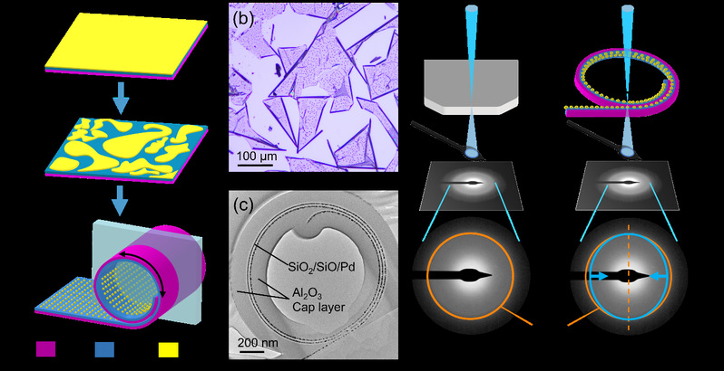
图1. 多层自卷曲纳米薄膜的样品制备和NBED 应变测量实验图示。 (a) 使用干法刻蚀释放和快速热退火处理制备样品的示意图。底部图示中的浅蓝色阴影部分说明了通过聚焦离子束(Focused ion beam, FIB)切割和制备透射电子显微(TEM)横截面样品的示意图。 (b) 自卷曲纳米薄膜样品的光学显微镜图像。 (c) FIB 截面样品的明场 TEM 图像。 (d) 和 (e) 分别是未受应变和受应变样品的 NBED 图案及应变示意图。
Figure 1 Illustration of sample preparation and NBED strain mapping of multilayer rolled-up nanomembranes. (a) Schematics of sample fabrication using dry-releasing rolling with rapid thermal annealing treatment. The light blue-shaded part in the bottom panel illustrates the FIB cutting for the cross-sectional TEM samples. (b) Optical microscope images of the rolled-up samples. (c) Bright field cross-sectional TEM image of a FIB fabricated sample. (d) and (e) The NBED experiment schematics of unstrained and strained samples, respectively.
研究团队首先分别通过传统湿法和颗粒辅助干法释放了自卷曲的非晶纳米薄膜,如图1 (a)-(c) 所示,由于非晶材料的无序性质,在 TEM 中无法直接获得原子位置信息或其对应于周期性排列的离散衍射点。而非晶材料的结构信息被编码在倒易空间的扩散衍射环中,可以通过空间分辨率小至1-2纳米的纳米束扫描样品的不同区域,生成衍射环并构建其各向异性与实空间结构信息的关系。图1 (d) 和 (e) 分别说明了没有残余应变和有残余应变的样品之间的倒易空间(衍射图案)的差异。结果显示局部切向应变方向与自卷曲纳米薄膜样品的几何形状之间的一致性。同时湿法和干法制备释放的两个样品中的残余应变显着不同,不仅是由于材料的选择不同,更重要的是由于金属纳米粒子的存在引起的应变增强。目前的结果证明了在具有复杂 3D 结构的非晶样品中进行纳米级应变测绘的可行性,为柔性电子器件中的高分辨率应变分布表征提供了解决方案,局部应变的定量测量可以更深入地了解纳米级膜的自卷曲过程。
As an important component of nanomembrane materials, amorphous nanomembranes play a crucial role in flexible electronics and other important applications due to their ability to create complex 3D structures through strain engineering. In order to better understand the formation of these structures, it is necessary to accurately measure and map the local elastic strain distribution with high precision at high spatial resolution, providing a path for the design and preparation of amorphous nanofilms with excellent properties.
On March 5, 2024, our institute published a paper titled Measurement of residual elastic strain in rolled-up amorphous nanomembranes using nanobeam electron diffraction in Applied Physics Letters. PhD candidate Zhi Zheng is the first author, and Professor Yongfeng Mei and Changlin Zheng are the corresponding authors. This work is strongly supported by the Department of Physics and the State Key Laboratory of Surface Physics of Fudan University, and the State Key Laboratory of Molecular Engineering of Polymers of Fudan University. In this study, the research team conducted nanobeam electron diffraction (NBED) studies in different directions on various rolled-up amorphous nanomembranes. By analyzing the diffused diffraction rings obtained at different positions on the amorphous sample, the anisotropic structural information in the reciprocal space was extracted and the local strain distribution in the real space was determined. Analysis shows that particle-assisted dry-etched and released samples exhibit higher local strain values than purely amorphous wet-etched and released samples. This suggests that the nanoparticles introduce additional strain through the dewetting effect, thus promoting the formation of self-rolling 3D structures.
The research team first released self-curling amorphous nanofilms through traditional wet methods and particle-assisted dry methods, as shown in Figure 1 (a)-(c). Due to the disordered nature of amorphous materials, the atomic position information or arranged discrete diffraction spots corresponding to periodicity cannot be directly observed in TEM. The structural information of amorphous materials is encoded in the diffusion diffraction ring in reciprocal space. Different areas of the sample can be scanned with a nanobeam at a spatial resolution as small as 1-2 nanometers to generate the diffraction ring and construct the relationship between anisotropy in reciprocal space and real space. Figure 1 (d) and (e) illustrate the differences in reciprocal space (diffraction pattern) between samples without and with residual strain, respectively. The results show consistency between the local tangential strain direction and the geometry of the self-rolling nanomembrane samples. The residual strains in the two samples released by the simultaneous wet and dry etching are significantly different, not only due to the different choice of materials, but more importantly due to the strain enhancement caused by the presence of metal nanoparticles. The current results demonstrate the feasibility of nanoscale strain mapping in amorphous samples with complex 3D structures, providing a solution for high-resolution strain distribution characterization in flexible electronic devices, and quantitative measurement of local strain, enabling further understanding of the self-rolling process of nanoscale membranes.
文章信息:Zhi Zheng, Chang Liu, Wenhao He, Jiayuan Huang, Jiachuo He, Gaoshan Huang, Yongfeng Mei, and Changlin Zheng. Measurement of residual elastic strain in rolled-up amorphous nanomembranes using nanobeam electron diffraction. Applied Physics Letters 124.10 (2024).
文章链接:https://doi.org/10.1063/5.0190880